|
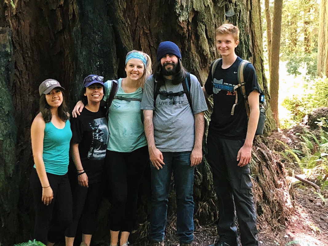
Hello, hello! This past week, our research proposals were due, meaning we all had to nail down our research questions and plan how we’ll work to answer them in the next six weeks. After a lot of thought, I’ve decided to investigate the feeding ability and morphological trade-offs of partial echinoderm larvae. When larvae are going about their lives in the ocean, they need to be able to collect food from their surroundings. Since algal cells are not particularly dense in ocean waters, it isn't efficient for larvae to just gulp up a bunch of water and filter it. Rather, they detect prey one-by-one and, ideally, capture and retain it. Central to this ability is the ciliary band – the principal feeding and swimming organ of a growing larva. In echinoderms, the ciliary band consists of a dense strip of ciliary cells that beat away from the mouth and, in response to detection of prey, produce local reversals of ciliary beat to redirect parcels of fluid toward the mouth (Strathmann 1975). Interestingly, purple sea urchins (Strongylocentrotus purpuratus) and bat stars (Patiria miniata), which have very small and large egg sizes respectively, are both able to produce functioning ciliary bands for feeding and swimming. I’m interested in understanding what happens to the ciliary band when a developing embryo loses a portion of this initial investment and must begin with a smaller starting point – will they still be able to produce a functioning ciliary band? If so, does this require other parts of the larva to compensate? Half- and quarter-sized embryos can develop into seemingly normal larvae. But what are the impacts of this manipulation on the ciliary band? One possibility is that the larva and all of its structures, including the ciliary band, scale down equally. Another possibility is that a partial larva would instead prioritize certain structures and cells over others, leading to an altered morphology. If the latter case is true, then the larva must compensate for these disproportionate structures, losing cells from certain areas to allow for a greater number of cells in others. By fluorescently tagging areas of interest, I hope to better understand the impacts of these embryological manipulations on the structure and function of the ciliary band in echinoderms. Next week, I’ll go more into the details of my project and how exactly I’ll be investigating these questions. In other news: Last weekend, a few interns and I made the trip down to California to do a little hiking and camping in the Redwoods! Our hike in Jedidiah Smith State Park was absolutely breathtaking. The first half of our hike consisted of a lot of silence interspersed with exclamations of amazement as we took in the nature around us. The way back went by a lot quicker, as we were (or at least I was) more preoccupied with the thought of s’mores awaiting us back at the campsite (did I mention I love s’mores?). On Tuesday, we discussed how to make scientific posters, and half of the REUs presented their research proposals. It's so great to hear about what everyone is doing and I'm so excited to see how everyone's projects turn out.
DOG SPOTTING!!!
0 Comments
Leave a Reply. |
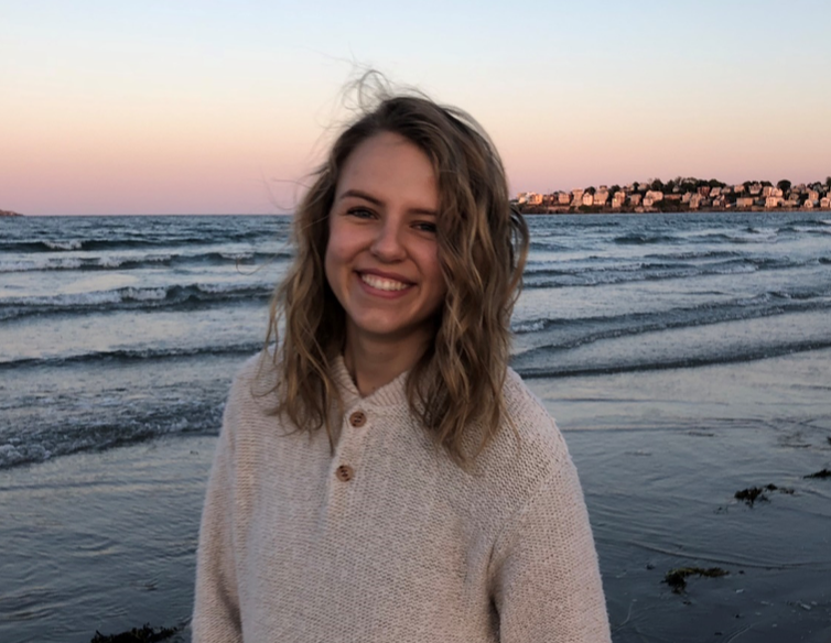
AuthorHello! My name is Ana and I am a rising senior studying biology and music at the College of the Holy Cross in Worcester, Massachusetts. This summer, I am working under the mentorship of George von Dassow. I am looking forward to seeing where my research takes me and to becoming a part of the OIMB community! Archives
August 2018
Categories |


 RSS Feed