|
Hello! My name is Nina Denne and I just finished my first week as an intern in the REU program at the Oregon Institute of Marine Biology (OIMB). I’m originally from the Chicago suburbs, but I go to Carleton College in Minnesota, where I am majoring in Biology with minors in Russian and Neuroscience. At Carleton, I do cell biology research focused on the bacterium Ralstonia solanacearum, which infects plant tissue and causes bacterial wilt disease. Although I’ve always been fascinated by marine biology, I’ve never had a chance to do any type of marine science before. In fact, I had never even seen the Pacific Ocean before this weekend!
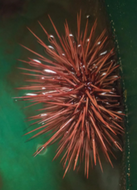
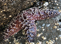
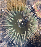
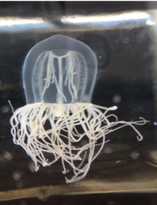
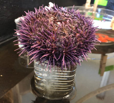
Above: Just a few of the organisms I’ve met this week. Left to right: a sea urchin, a sea star, an anemone, and a jellyfish. At OIMB, I am working in Dr. George von Dassow’s lab. His research focuses on embryology of marine invertebrates including sea urchins, sand dollars, jellyfish, sea stars, and others. Specifically, current research questions involve the role of proteins Ect2 and Rho in the organization of actin (a part of the cell’s cytoskeleton) during cytokinesis (cell division). However, prior to delving into the research, we needed to develop some technical skills. One of the first skills we tackled was injecting developing embryos with probes containing instructions to make green fluorescent protein (GFP) fused to a protein of interest. Additionally, we practiced melting and reshaping glass tubes to form a curved tiny glass tube that will let in a single embryo at a time. This glass tube is connected to rubber tubing with a mouthpiece on the other end; this is essential because transferring embryos requires high precision, and mouth-eye coordination is more precise than hand-eye coordination.
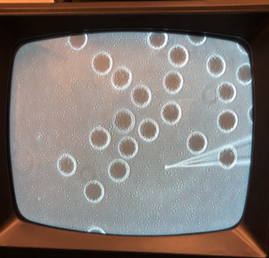
Left: A purple sea urchin releasing its eggs. Right: Injection of recently fertilized sea urchin eggs with a red fluorescent probe. Since most of Dr. von Dassow’s work involves microscopy and working with gametes and embryos, we also tried looking at a variety of subjects under different microscopes for practice. The subjects included sea urchin embryos, sea star oocytes, jellyfish oocytes, and more. One of the subjects I was most excited to work with was the diatom – a single celled alga with a silicon cell wall. Although I had worked with diatom genomes in a previous lab at the Chicago Botanic Garden, I never met them. This week, I was able to see a diverse array of diatoms under the microscope, collected from a plankton tow. In addition to diatoms, I also met some dinoflagellates, a group of protists found in plankton that have a single tail (called a flagellum). The species that Dr. von Dassow keeps as a pet has another interesting quality: they are bioluminescent, meaning they glow in the dark when slightly agitated. On Wednesday evening, I came back to lab in the evening to see the light show produced by the dinoflagellate Pyrocystis and was amazed by waves of glowing green specks!
0 Comments
Leave a Reply. |
AuthorI am a rising junior at Carleton College, majoring in biology with minors in Neuroscience and Russian. I'm very excited to be working in Dr. George von Dassow's lab this summer, where I will be studying cell biology and embryology of marine invertebrates. Archives
August 2019
Categories |
 RSS Feed