|
This has been an incredible first week. Each day presents new, challenging, information and lab techniques to learn. My mentor, George von Dassow, has blown my mind several times a day. My lab mates, Nina Denne and Sadie Rose are wonderful. The very first thing we did was walk down to the ocean and catch jellyfish right off the docks. George told us about how green fluorescent protein (GFP) was originally isolated from the Aequorea victoria jellyfish that we had collected in a bucket next. The use of GFP has since transformed the field of cell biology with big implications for many fields including medicinal imaging. Back in the lab we are learning lots about microscopy and embryology. As a way of practicing taking photographs with the microscope, I set myself the task of photographing the early embryonic stages of the purple sea urchin (Strongylocentrotus purpuratus). George recounted that he was once carrying a sea urchin from one side of the lab to the other when he got caught up in talking to someone in the hall. Without thinking about it be started to bounce the urchin up and down in his hand. To his surprise, when he looked down, it had spawned! What a happy accident! Using the now famous shake technique, I was able to get the urchin to produce eggs. Ready for use, they can be combined with sperm cells to produce zygotes. Once a sperm enters an egg the fertilization envelope develops as a block to polyspermy, the fertilization of one egg by multiple sperm. Watching the moment of fertilization happen under the microscope blew my mind. Of course I had seen video of the event, but knowing it was happening right then, underneath my lens, was something I will never forget. 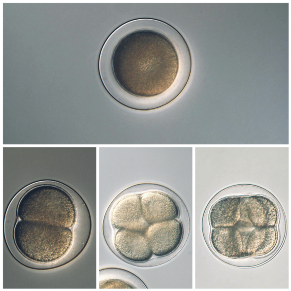 You can see the process of early development in the sequence above As of this writing, besides the sea urchin (Strongylocentrotus purpuratus), we have collected or fertilized embryos of a jellyfish (Aequorea victoria), a sea star (Patiria miniata), a sea snail (Calliostoma ligatum), acorn barnacles (Balanus glandula), and gooseneck barnacles (Pollicipes polymerus). I’ve also started to keep a few pets including some nudibranchs, sea cucumbers, and bioluminescent dinoflagellates. Marine invertebrates are a pretty diverse bunch! Stay tuned next week for photographs of the continuing development of the purple sea urchin embryos. This is what they looked like at the end of this week: 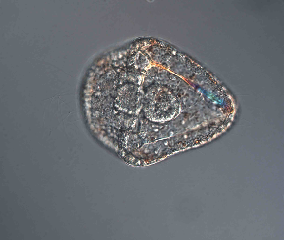 Next week we will begin to define our summer research projects which will almost certainly involve working with eggs, embryos, or larvae of various invertebrates and some combination of fluorescence, time-lapse, and confocal microscopy.
See you next week!
0 Comments
Leave a Reply. |
AuthorMy name is Philip Aspinall, and I am a student at Sierra College in Grass Valley, California. The first time I peered into a microscope and found an entire, complex, beautiful world below the visible, I was transfixed. I am thankful for George von Dassow and Svetlana Maslakova for allowing me to work in their lab, and to Geroge for his generosity with his time and for being my mentor this summer. Archives
August 2019
Categories |
 RSS Feed