|
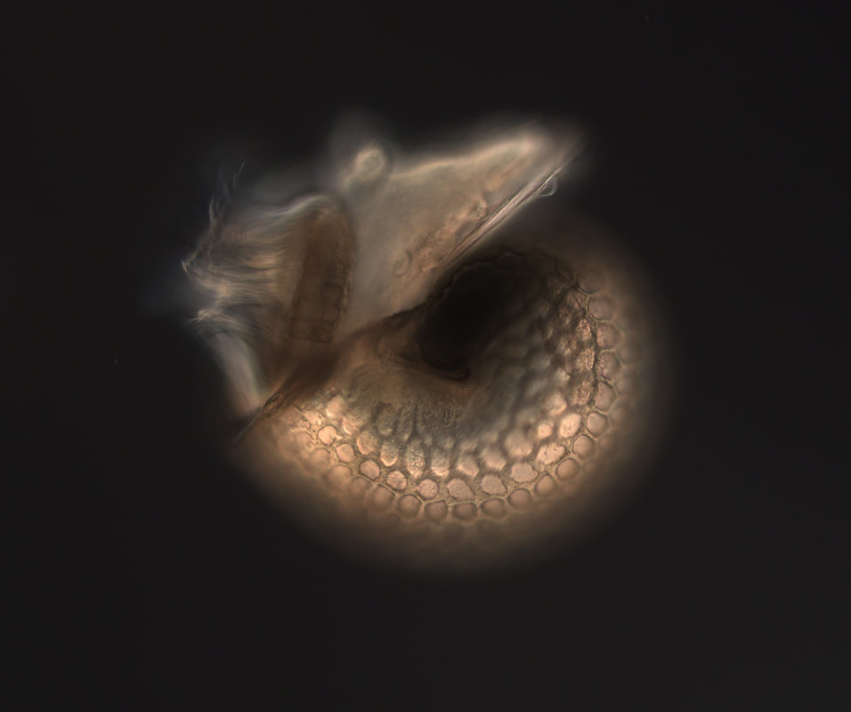
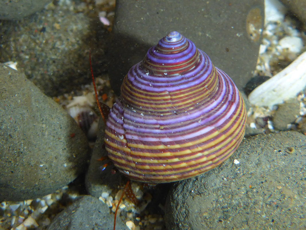
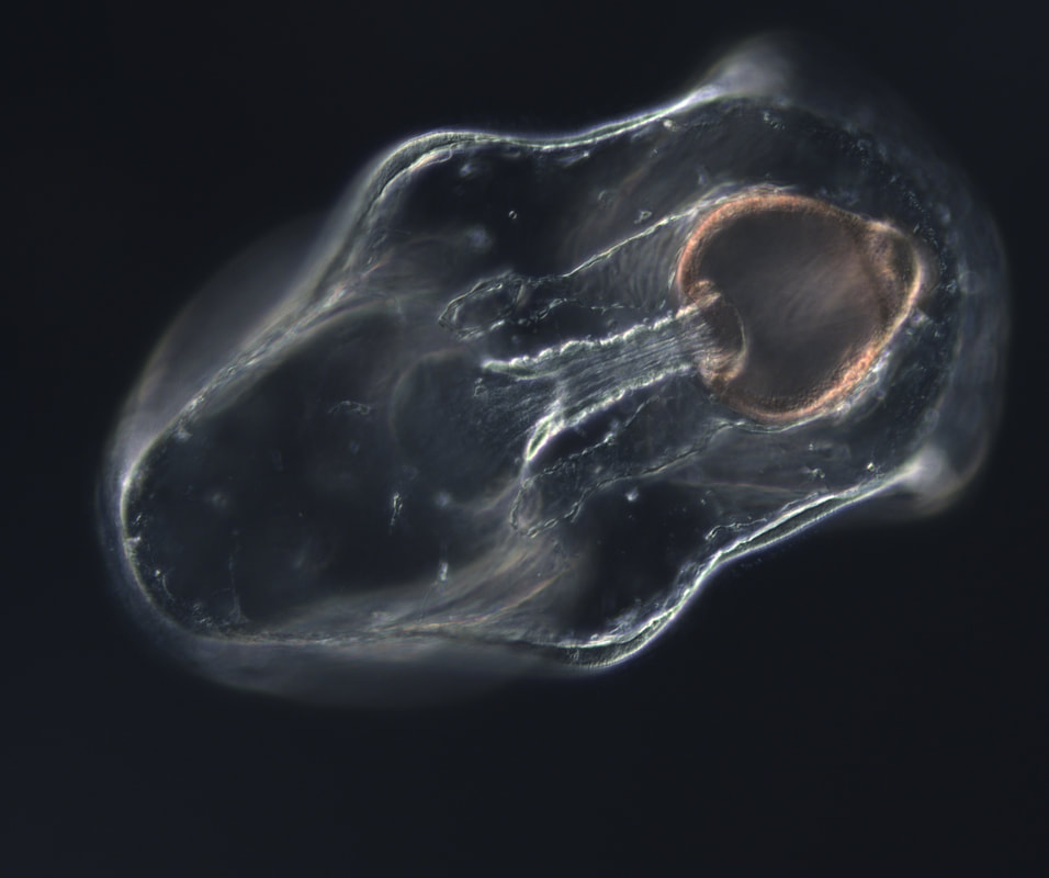
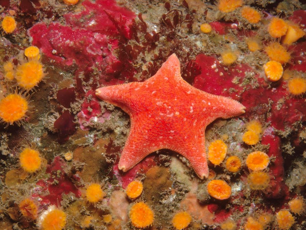
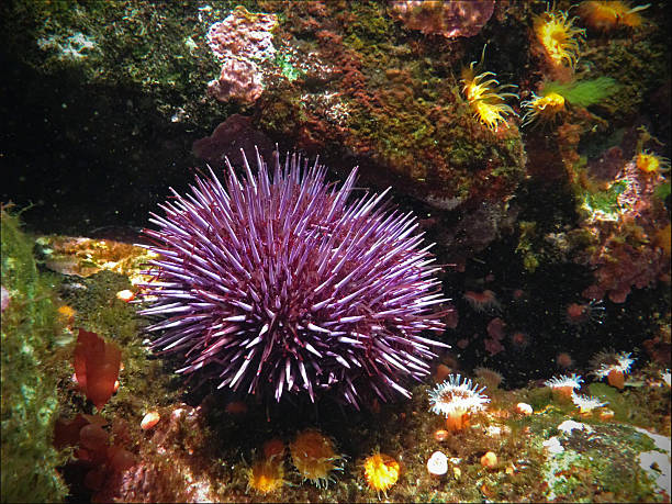
So far the goal has been to collect wild jellyfish, and keep them on the sea tables in the lab. Then to use a recently discovered HORMONE that induces them to spawn, collect the eggs, micro inject them with RNA that will target specific parts of the cell, making them fluorescent so that we can record them under the confocal microscope. The reason this is interesting, if just the process itself is not interesting enough, is that many jellyfish cells divide in a way that is different than other animals. The way that cells divide is only just beginning to be understood. George has been researching how a cell actually manages to coordinate its own division. What are the controls? The feedback loops? Maybe studying the mechanism in jellyfish, who essentially do it differently with the same equipment, will help us understand more about the mechanism in general. If you know the basics of the cell cycle, then you know that cytokineses, the term for the dividing of the cell into two, normally happens in a ‘pinch’ like motion. The video below, taken by my mentor George von Dassow, shows a 'typical' cleavage pattern. Most jellyfish on the other hand divide unilaterally, starting on one side and moving to the other in a ‘zipper’ like motion. This video, also taken by George von Dassow, shows the early divisions of Phialidium gregarium, one of the species I am working with. So far it has been challenging to have a reliable supply of animals, as collecting them is a little boom or bust, they either are totally absent, or are there in great numbers. However, slowly but surely, I am feeling more confident knowing my way around the lab. I am getting the hang of which tides to go out looking for jellyfish, and the timing of getting them to spawn. The next important step is to successfully inject some eggs with the RNA probes and have them develop. Wish me luck! Outside of the lab I have been enjoying the beautiful, remote Oregon coast. I have been going out surfing at Bastendorff beach. All the other REUs are really nice and we had a perfect sunny day at the beach for 4th of July with a lot of the faculty, grad students, and University of Oregon students, eating great food, playing frisbee and even swimming. Last weekend we visited the main University of Oregon campus in Eugene to meet students doing summer research there. This weekend those students will be coming up to go camping with us and we will give them a tour of our lab.  So if you are playing along from last week with 'Guess that larvae' here are the answers. The pictures on the left are the microscopic larvae, the pictures on the right are what they become. Drum roll please. A) A snai! Also known as Calliostoma ligatum, commonly known as blue top snail, is a pretty little snail found in the inter tidal. B) A sea star! Also know as Patiria miniata, or the bat star. C) A sea urchin! Also known as Strongylocentrotus purpuratus, or the purple sea urchin.
0 Comments
Leave a Reply. |
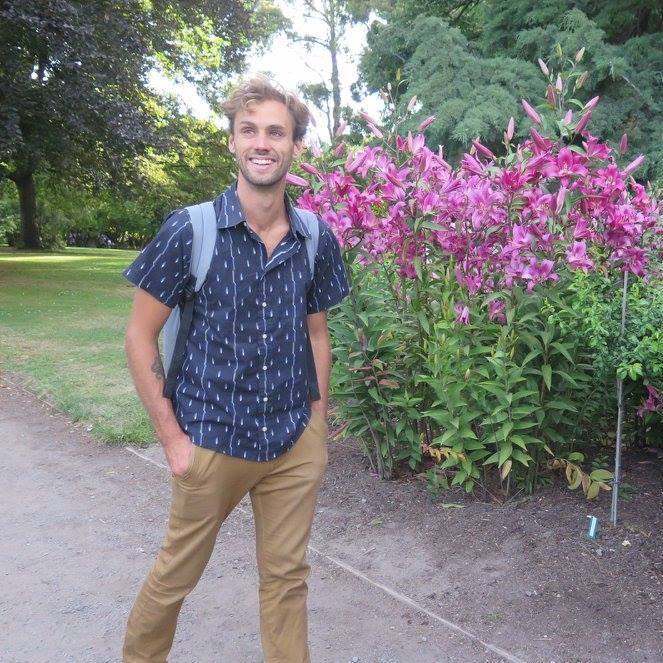
AuthorMy name is Philip Aspinall, and I am a student at Sierra College in Grass Valley, California. The first time I peered into a microscope and found an entire, complex, beautiful world below the visible, I was transfixed. I am thankful for George von Dassow and Svetlana Maslakova for allowing me to work in their lab, and to Geroge for his generosity with his time and for being my mentor this summer. Archives
August 2019
Categories |

 RSS Feed