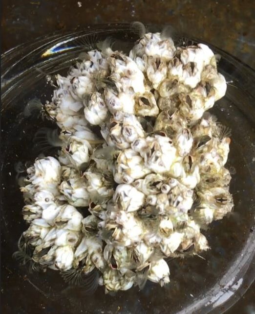
 Balanus glandula Balanus glandula Hi everyone! This is week two and I’ve got a project! On Monday Dr. von Dassow, my lab mates, and I discussed what we would be doing for our projects. I will be working with barnacles; Balanus glandula is their scientific name but informally they are called acorn barnacles. They are the most numerous barnacles on the Oregon coast and usually grow on docks and rocks. I will be studying cell polarization in barnacle embryos. In C. elegans cell polarization happens early, during first cleavage. When an egg is fertilized it starts dividing to create new cells. When the egg makes its first division it pushes some types of proteins into one half of the cell and other proteins into the other half of the cell. So, when the cell divides into two cells, one cell has a lot of some proteins and the other cell has a lot of other proteins. This happens because different parts of the creature will be formed from each cell. For example, part of one of the cells will eventually form the gastric tract. This cell will take the proteins that it will need to make this system. The moving around of substances is called cell polarization. I will be trying to discover if cell polarization happens in basically the same way in Balanus glandula as is does in C. elegans. To do that I will be using a probe to track atypical Protein Kinase C. The aPKC is found in many different organisms so there is a good probability that it will be found in Balanus glandula. I will be using a probe to track this protein and see if it will be doing anything interesting in barnacle eggs. Right now, I am practicing injecting substances into barnacle embryos and learning how to use the confocal microscope. This week I attached a video so I could show you what the barnacle embryos look like after a successful injection of RNA. What you will see is a bunch of white lines “flowing” through the embryos. The probe that was injected is a protein that sticks to the front of forming microtubules. When the microtubules stop growing, they either attach to a solid object or catastrophe which means it breaks apart. The video is sped up and is made up of pictures taken at different depths in the embryo. Helping to take this video was my introduction to working with the confocal microscope. If you watch the video closely you can see the cells dividing. On Sunday a group of the Interns went to Bastendorff beach. It was warm enough that we were wearing shorts and t-shirts! I know that I also enjoyed our meeting on Wednesday when we all gave a 5-minute talk on what our projects would be on. It was interesting to learn about everyone’s plans in more detail than just snippets from mealtime discussion.
0 Comments
Leave a Reply. |
AuthorHi! My name is Sadie and I just graduated from Central Oregon Community College in Bend Oregon. I am working in Dr. von Dassow’s lab and I am excited to learn about research and cells. Archives
August 2019
Categories |
||
 RSS Feed