|
Welcome to the week 8 post everyone! This week we’ve been doing a lot of work on our posters. On Tuesday we had a poster critique among the interns and Dr. Watts. We projected everyone’s posters up on the screen one at a time and gave feedback on what we liked, or thought could be changed. I greatly appreciated the advice. It was fascinating to see everyone’s posters taking shape. Then we turned the posters in for the final review on Friday. It has been surprisingly difficult to cram everything I have seen and observed onto one poster. The most constant comment I got at the critique was to get rid of a lot of the text! Trying to condense everything has made me sift through what I have observed and try to pick out the most important things. Probably the most important finding that we observed was a secondary cleavage furrow. The first and main cleavage furrow starts out like it is going to divide the embryo into two equally sized cells. Then a second furrow forms on the side and continues moving towards the center until it connects to the main cleavage furrow. The result is that there is one large cell and one small cell. The other thing I’ve been doing is sorting through videos and pictures. Dr. von Dassow taught me how to use some basic functions with ImageJ Fiji. The program is incredible, and I’ve had fun turning different probes into different colors to emphasize different pieces of the embryo. Normally we work with grayscale images because that is the best color range to pick out details in. Greens also work well but blue doesn’t show details at all. And while some color combinations, like green and magenta, clash they actually show off the different parts to the maximum advantage. I’m going to attach some images so everyone can see the difference. While I have enjoyed getting to look through the data and explore ImageJ I’m always glad when I have time to inject and image more embryos. Since I have all the data, I need I have been able to some new ways of setting things up and see what else I can learn with out the pressure of getting a result. On Friday we tried putting the embryos in a chemical that keeps their egg shell soft and pliable. Unfortunately it is a lot harder to get the embryos to stick to the injection plate while using 3 APA but we’re still working on it. Below you can see the pictures of the embryos, they are all the same brightness but you can see the details on the cell surface much better in the gray than the blue. 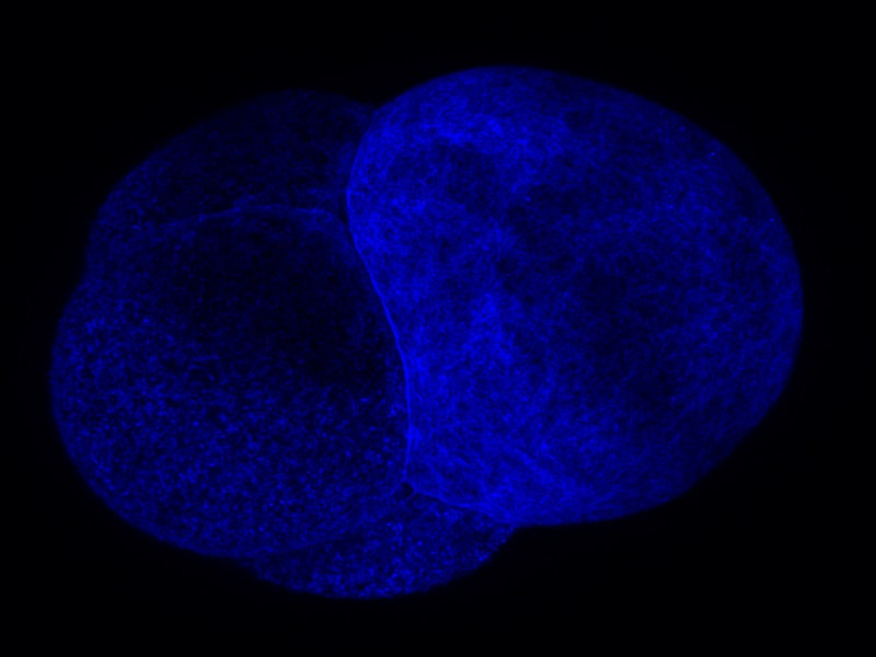 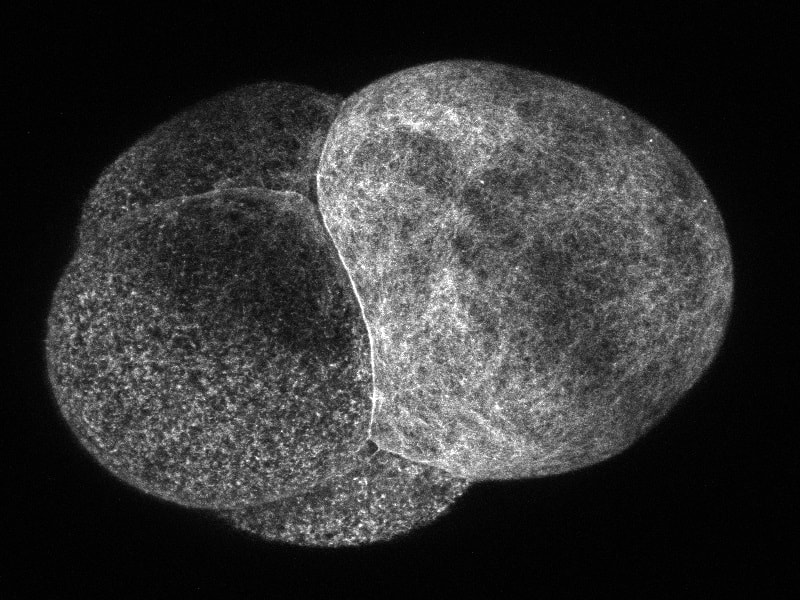 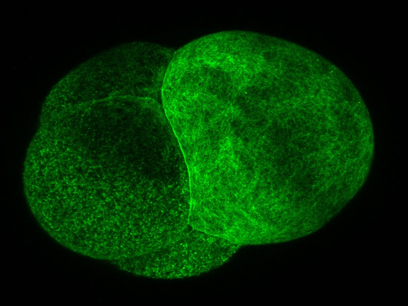
0 Comments
Leave a Reply. |
AuthorHi! My name is Sadie and I just graduated from Central Oregon Community College in Bend Oregon. I am working in Dr. von Dassow’s lab and I am excited to learn about research and cells. Archives
August 2019
Categories |
 RSS Feed