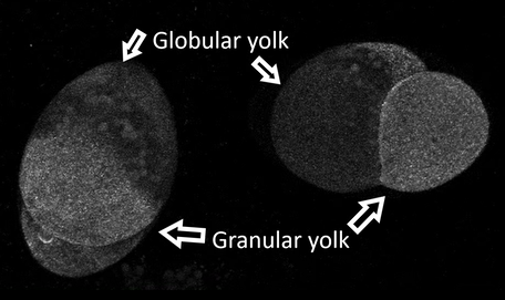
 The Differentiated Yolks The Differentiated Yolks Welcome to the week 6 blog post! Because the aPKC probe I was originally planning to use didn’t work, I have been working my way through a few other probes. I need to use a probe that allows me to see the structure of the cell and the cleavage furrow. On Monday, I tried two different kinds of myosin probes. Interestingly one of these probes was formed by using antibodies from camels! Although one of the probes was visible, it wasn’t particularly bright or very helpful. So, on Tuesday we tried a utrophin probe. The probe has been used before in the lab so we knew it would work. The probe lets us see the cleavage furrow, and if the spindle poles are close to the surface, we can see them too. We have been trying to get videos of the embryo dividing during first cleavage, but the success rate is very low right now. Let me rephrase that. We have plenty of videos of the embryos dividing, you just can't see the cleavage furrow because the embryo rotated around to hide the division! If I can see the spindle poles I can guess where the cell will divide. However, in barnacle embryos the first division is not symmetrical. The embryo divides into two cells; the first is small and contains the granular yolk and the second is large and contains a little of the granular yolk but also contains all the globular yolk. In the picture I have pointed out the two types, but you can also see the different sizes of the cells. So even though we typically know where the spindle poles are, we don’t know if the top quarter of the cell will become the smaller cell or the bottom quarter. So far it has been frustratingly hard to find a cell the divides where we can see the cleavage furrow. So, part of what I am doing now is looking at videos that have been taken and taking pictures of embryos in different positions so hopefully we can find a pattern that will let us choose the best embryos to view. While the video can only capture what is happening on and right below the surface of the side of the embryo closest the camera, the pictures let us see about half way through the embryo. The picture below is one of the ones I took on Thursday using the utrophin probe. 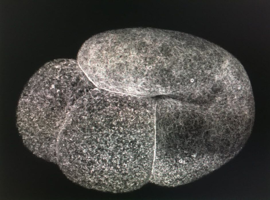 The Utrophin Probe
0 Comments
Leave a Reply. |
AuthorHi! My name is Sadie and I just graduated from Central Oregon Community College in Bend Oregon. I am working in Dr. von Dassow’s lab and I am excited to learn about research and cells. Archives
August 2019
Categories |
 RSS Feed