|
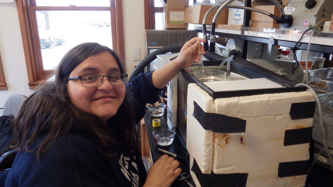
Hello everyone! It’s been about a week since my last blog post, and a lot has happened. Over the weekend, Richard, our sea captain Knute, and Richard’s graduate students Nicole and MacKenna took all the REU students on a trip to dredge for sea creatures on the R/V Pluteus. We started our journey early in the morning and the ride was rough and nauseating for practically everyone on board. After some time, and a lot of fortitude, we were able to collect many different fascinating creatures! We collected some sea cucumbers and peanut worms, enough to start a big and slimy salad! We also found basket stars, various crab and shrimp species, and some cockles. The dredging was hard work, but ultimately an exciting experience! The ride back to shore was even more nauseating, but after some fresh air, and an even fresher slaps of seawater to the face, I managed to keep down my breakfast. 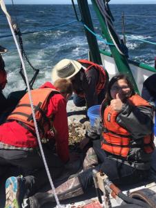 Smiling through the nausea! Smiling through the nausea! During my week in the lab, I had the main goal of practicing and perfecting my proposed methods of research, as well as prepare to write my proposal. At this time, I found that to study the functional morphology of the furcal rami, I needed to understand how to induce swimming behavior in my study cyprids. After doing some research using Eleanor Lamont’s master’s thesis, I learned that I can make cyprid larvae swim by manipulating light. Cyprids display positive phototaxis, which means that they have a behavioral tendency to swim towards sources of light. So, if I shine light upon my study animal, I can easily encourage them to swim for my observations. Next, I wanted to see how feasible it is to remove the furcal rami and still have the organism live from the taxing surgery. I conducted trial-upon-trial and eventually I decided that I currently do not have the tools or skills needed to remove the tiny furcal rami from the animal. What I could do, however, is safely remove the setae from that area of the animal and revive it. It takes an extremely steady hand to conduct this microsurgery. Its going to take many more attempts to remove setae, followed by revivals, to determine the feasibility of this procedure. If I can continuously remove the setae from the animals, then I will film their swimming behaviors with a high-speed video camera. A standard microscope camera (30 frames/s) is not quick enough to resolve the extremely fast thoracic appendages and the furcal rami. Finally, during my week in the lab, I attempted to study the musculature found within the caudal rami of the cyprid larva. The first try was simple. We removed the entire thorax from the organism and examined it on a microscope under polarized light. After a while of fiddling around with the settings, Richard and I saw some muscle bands leading from the thorax to the caudal rami. After confirming the presence of the muscle bands, we decided to try to stain the creature to see the neurons of the thorax. We used a common staining chemical called methylene blue in order highlight the neurons found within the thorax of the cyprid. Staining is an imprecise science, it’s quite difficult to gauge how long an animal, or part of an animal, should sit in the solution for full staining power. After quite a bit of trial and error, I figured that letting the thorax of the cyprid soak for four hours was enough time for the dye to bind to the neurons. However, after all that trial and error, I still couldn’t see the nerves well enough. So, my new plan is to remove the limbs individually and try to see their nerves. Science is all about the process of failing millions of times until you get the one right solution. All I can do is focus on the small victories and try harder next time. So onward to next week! 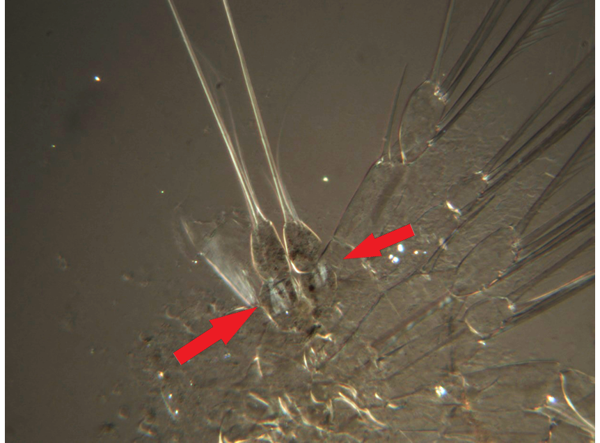 The arrows on the photograph are pointing to the muscle bands found within the caudal rami of the cyprid thorax. The abdomen appears to be squashed quite a bit, but the muscles were found at the segment closest to the body.
0 Comments
Leave a Reply. |
AuthorHello, I am Savanna Cabrera, a fourth-year zoology major from the University of Florida studying barnacles. I’m an avid arthropod admirer. Archives
August 2018
Categories |
 RSS Feed