|
We are officially halfway through the program!!! Kostantina and I have become more independent in lab, which means preparing cultures of half- and quarter-sized larvae on our own. This also means that there is a lot more trial and error and a short life expectancy for many unlucky embryos :( Some blastomere separations go smoothly, only to have the culture decimated by some evil bacteria a couple days later. Some seem to be going smoothly and then at the very end things don’t work out. Some don’t go smoothly at all in the first place. But that’s research I guess! It can be frustrating at times, especially given that time is limited, but when things do work it’s really rewarding. 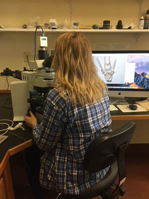 Looking at a happy and healthy sand dollar larva Last week I promised I’d go more into the details of my project and what exactly I’ll be doing in lab, so here I am! In order to understand the impacts of scaling on the functionality of the ciliary band, I also want to take a look at the effects of scaling on the larval nervous system. The ciliary band is the center of the larval nervous system and nerve cells are innervated throughout the ciliary band. So, let’s say that when we “shrink” down these larvae into halves or quarters, larval structures don’t scale proportionately and the larva appears to prioritize certain structures – like the ciliary band that is oh-so essential for feeding and swimming – over others. If this is the case, then other parts of the larva will need to compensate. Since the ciliary band and nervous system are so closely associated, it is possible that nerve cells would end up as one of the ones compensating. The loss of these nerve cells, which appear to play an important role in sensing and detecting prey, could end up reducing the functionality of the ciliary band. On the other hand, if there is proportionate scaling, it’s possible that the density of ciliary band cells will be so reduced that there aren’t enough cilia to effectively detect and capture prey. In order to observe ciliary band density, we’ll be using a fluorescent tag (GFP-centrin) that labels basal bodies, which are organelles found at the base of ciliated cells. This involves microinjecting eggs, a process which essentially consists of injecting eggs with a very tiny needle filled with RNA encoding a given fluorescent marker. We can then use a confocal microscope to compare ciliary band density in whole, half-, and quarter-sized larvae. In short, the confocal microscope is a microscope that eliminates all out-of-focus light and creates a 3D image of a fluorescently labeled specimen. To visualize nerve cells, we’ll stain larvae with a neuron-specific antibody called 1E11 and observe again with the confocal. Ideally, I’d like to get images of the ciliary band and nervous system for whole, half-, and quarter-sized larvae of sea urchins, sand dollars, and starfish. This will mostly depend on our blastomere-separating and culturing skills, so here’s to hoping things go a little better in this coming week! If time allows it, I’d also like to observe larvae using high-speed video to see what the impacts of scaling are on their ability to capture escape-prone prey.  Kostantina and George looking at a half-sized sand dollar larva using the confocal microscope
0 Comments
Leave a Reply. |

AuthorHello! My name is Ana and I am a rising senior studying biology and music at the College of the Holy Cross in Worcester, Massachusetts. This summer, I am working under the mentorship of George von Dassow. I am looking forward to seeing where my research takes me and to becoming a part of the OIMB community! Archives
August 2018
Categories |
 RSS Feed