|
We rise early and sleep early at the research station, stepping out in the mornings onto the fog-banked marina. Egrets, cormorants, and pelicans, commuters from across the bay, remind me that the sea has edges. At the start of the day, I like to walk across the bridge and back, or to visit the lookout point we found in the woods.
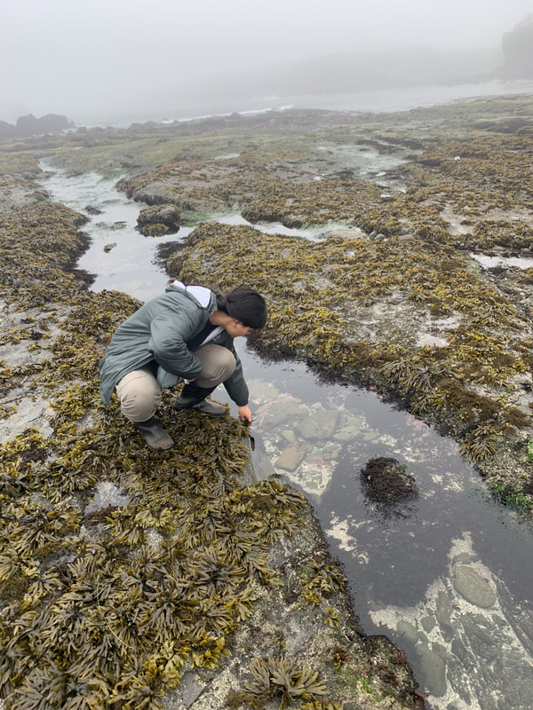
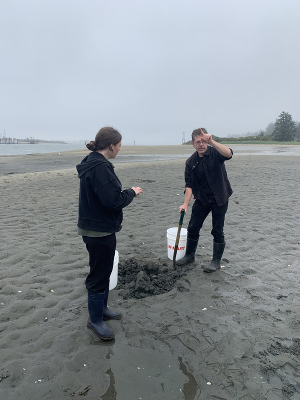
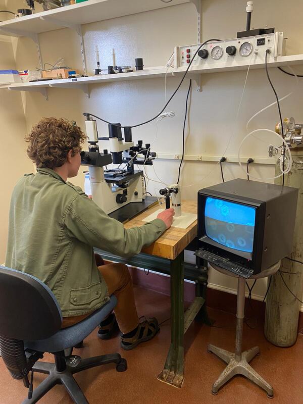
This week saw the beginnings of our projects. Chloe and I first slopped down onto the mudflats to dig up buried worms - the parents of her larval subject - and antagonize shore crabs; then we harvested sea star eggs and learned to operate the microinjector, which will be one of my main tools. For my project, I want to examine a peculiarity of cell division in sea star eggs. Cell division is a dynamic process, but it relies on a basic series of steps. Imagine that I need to bisect a circle of paper. First, I’d use a ruler and compass to orient myself with the center. Then I'd take a pen and draw a dotted guideline there. Finally, I’d fold and cut along the line. Simple as that. Animal cells, as it turns out, go about things in a similar way, using proteins. They have a folding machinery (called actin, also responsible for muscle contraction), an “inked” guideline (called Rho), a drawing hand (called Ect2), and a compass to direct it (the thready spindle structure of mitosis). But from a protein’s perspective, a cell is not a neat geometry on paper: rather a crowded, whirring industrial city. Just look at this section of the cellular landscape modeled by Evan Ingersoll & Gaël McGill. 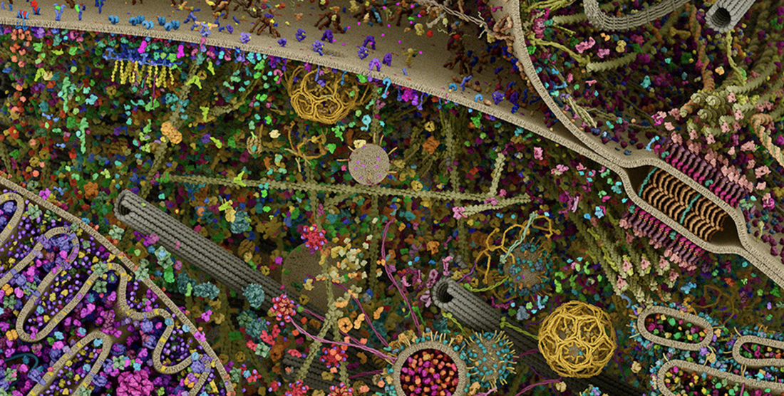 Down here, there are no straight edges, no convenient blank spaces to draw in, and nobody looking down to guide it all. Our sequence of steps, communicating only by touch, must coordinate with each other to act in synchrony across that entire city. A sea star egg cell measures about a quarter of a millimeter across - large enough for the human eye to see. It is a very big, very busy city, and sometimes the signals, the steps of division, get confused. Yet these first divisions touch off the entire undertaking of animal life. An invertebrate egg, cast out into the sea, must be especially keen to get itself divided and functional. What’s a cell to do? For the past few years, George’s lab has probed the way Ect2, Rho, and actin work together in sea star eggs. By tweaking their sensitivity, we can spark off striking ripples of activity at the cell membrane, causing Rho and actin to assemble and fall apart again in waves. It seems to be an extreme expression of a natural process, a feedback loop built into the cell. But what’s it for? Well, here’s one idea: how about an amplifier to help the machinery of division communicate across a big city?
Rho waves on the surface of a sea star egg with its excitability turned up through excess Ect2 protein. This figure was published in Nature in 2015 ( Bement et al ).
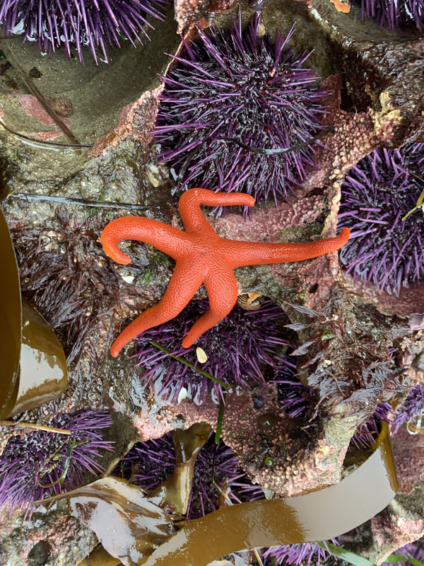
Hopefully, I’ll be able to test this idea in my project this summer. We might take a sea star egg and chemically nudge Ect2 and Rho a little, then watch how the subsequent divisions go. The difficulty lies in catching it on camera… and in doing this without killing them! I wanted to practice on the fertilized eggs of the ochre star, Pisaster, a common sight in the tidepools. But after severely traumatizing nearly a dozen Pisaster specimens (gamete harvest for this species involves lopping off an arm) and finding not a single fertile male, we got fed up. We’ll be using the bat star Patiria miniata for our experiments instead. Expect to see a lot of pictures of Patiria eggs in the coming entries. Lots and lots...
0 Comments
Leave a Reply. |
|
 RSS Feed