|
Things are starting to work a little better around here. I have switched to keeping the jellyfish in an environmental chamber so they are always at 12 degrees celsius and setting a regular feeding schedule of Artemia (brine shrimp) nauplii means the jellies are much happier and more cooperative in the lab. This means that my workflow in the lab has become more regular and I can depend on getting fertilized eggs within a certain window of time. I have even gotten some of the planula, the larval stage of the jellyfish, to settle and form hydroids, the immobile and colonial phase. See my last post for a review of jellyfish life cycles.
All of our experiments work better with fresh animals. I read today that Phialidium gregarium, “particularly during late spring and summer, this tiny jelly is so abundant as to appear to be filling the ocean.” This year, for whatever reason, this has not been the case. Next week we will get to present our research to the public at the Charleston Marine Life Center (CMLC), a natural history museum that serves as the Oregon Institute for Marine Biology’s public outreach. It should be fun to come up with a way to show my research to a general audience. Often when I go looking for jellyfish there are families fishing or crabbing from the docks and inevitably the kids (and the adults) get curious about the jellyfish. So, I guess I’ve already started getting some practice communicating my science to non scientists.
0 Comments
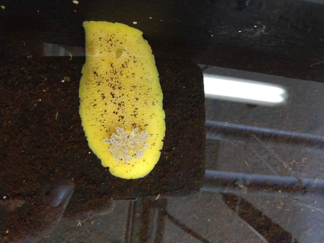
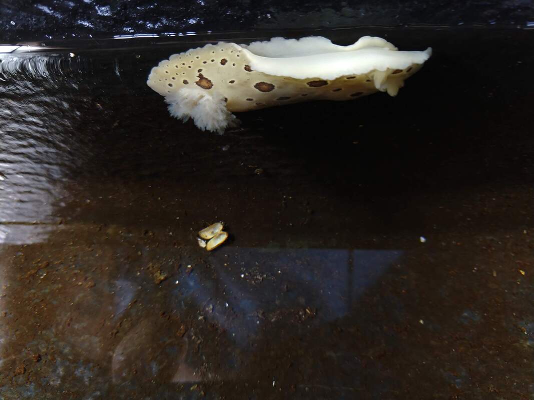
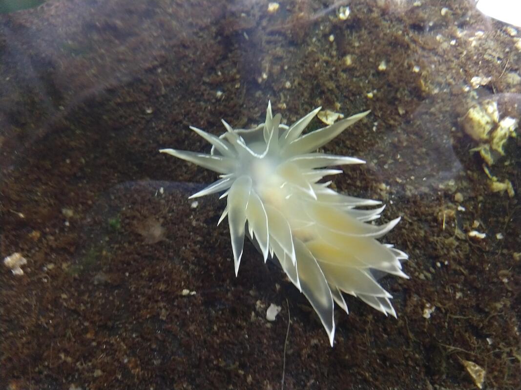
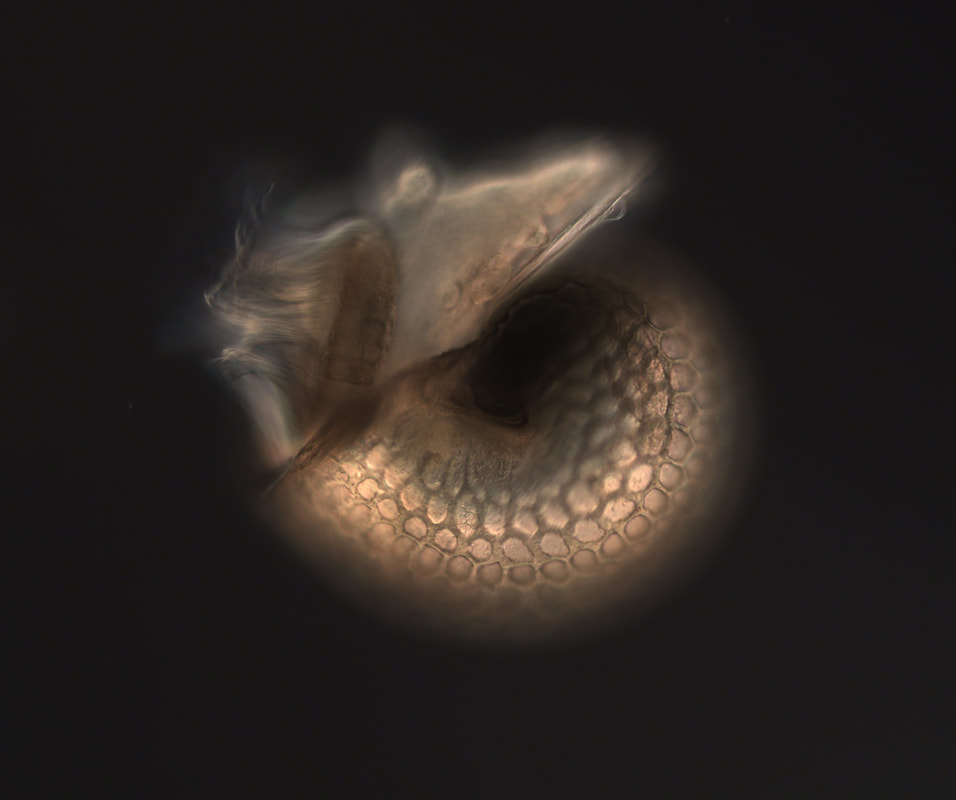
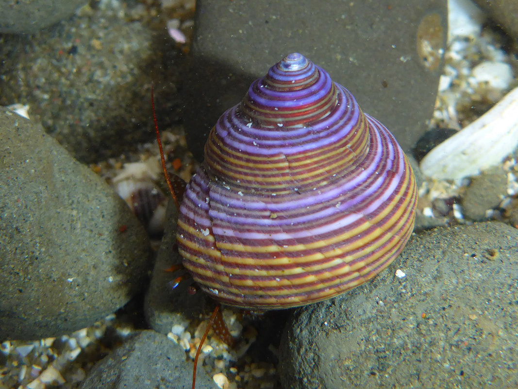
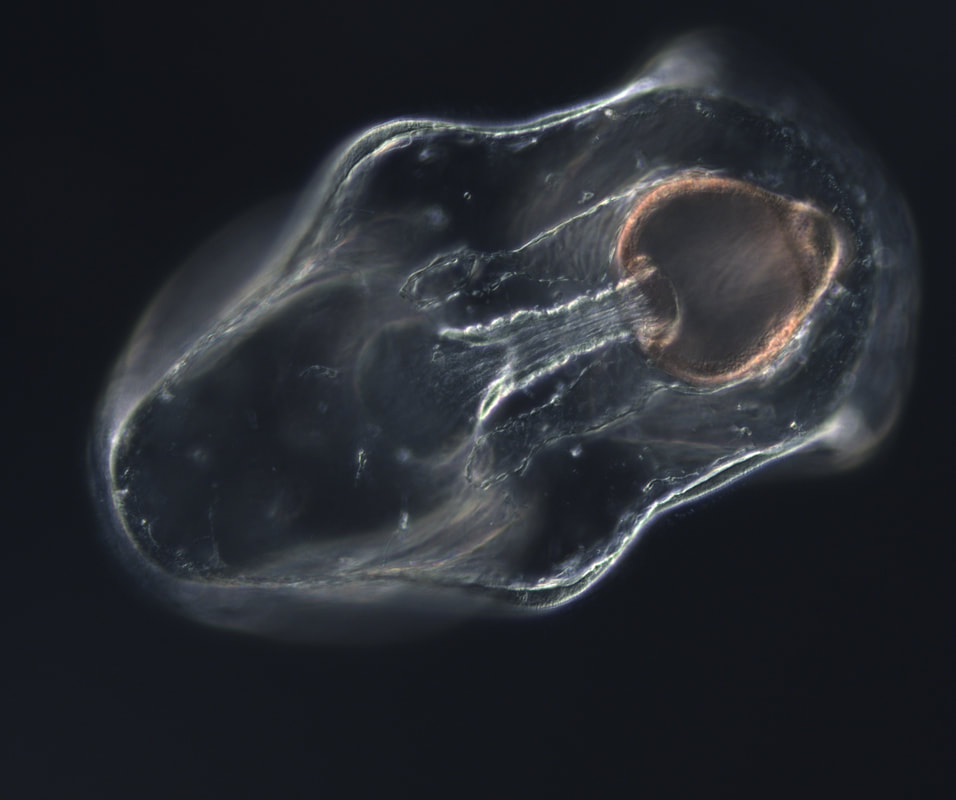
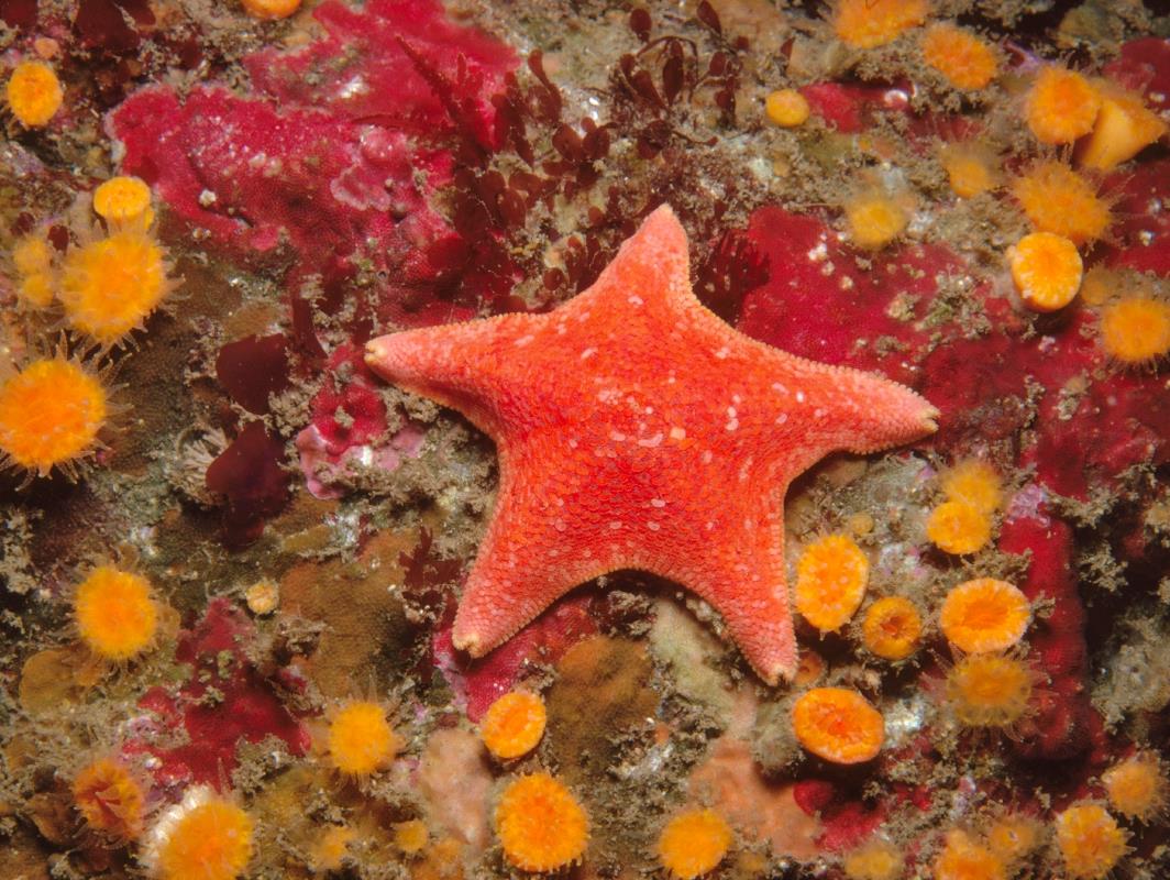
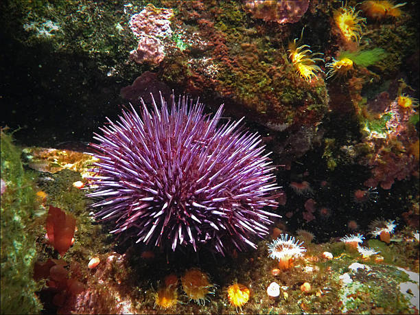
Incredibly another week has gone by. Work in the lab coming along but not much has changed from last week. We are still working on getting conditions right for the jellyfish. In the mean time, I thought it would be interesting for you to learn a little bit about the hydrozoan jellyfish's life cycle. There is more to it than you might know. The life cycle of the species I am studying, Phialidium gregarium, is typical of hydrozoans. The free swimming medusa, what you think of when you think jellyfish, is the sexually reproducing phase. Individuals produce have either ovaries or testes (although this week we found a hermaphroditic individual that had two ovaries and two testes). Light cues induce the medusa send sperm and eggs into the water. Fertilization is external and the embryo develops into a small larva called a planula. Now this is where it gets interesting. After some time swimming around as a non feeding planula, the jellyfish settles down and metamorphoses into a polyp. The polyp has a stalk and tentacles around its mouth, and it somewhat resembles a tall sea anemone. The polyp reproduces asexually by cloning itself and becoming a colony of polyps. After a while the colony will produce a specialized polyps that release medusae. Once the medusa mature, the cycle starts again. 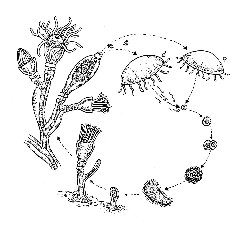 Hello again dear reader, Another week has passed and we continue to have trouble with the jellyfish being reliable. There was however a bright glimmer of hope as we actually took our first movie of unilateral cell division in jellyfish embryos on the confocal microscope this week! This is what we have been waiting for. When we finally got the dividing cells on the microscope everyone in the lab was so excited that four of us missed lunch to watch. It was really fun (and someone saved us all a plate). The cells had been injected with ‘life act’, a probe for actin, and the movie we captured does show us some very promising features. Mostly, the famous and elusive contractile ring was nowhere to be seen. Which is interesting because if the contractile ring model is not the whole picture, how are these cells dividing? We hope to get some more jellyfish soon and try it again. Ideally we will take more videos, with probes for different things and from different angles. I knew coming into this project that it was going to be ‘exploratory’ science with no guarantee of success and I am starting to come to terms with that. At the same time, I really hope it works! Stay tuned to see if the jellyfish come back next week In the mean time, I'd like to introduce you to seven nudibranchs that we have found on the docks when we go down looking for jellies. Check them out! We also looked at some bryozoans and ascidians under the stereo microscopes. Both of these were types of tiny, beautiful colonial animals that you would never see if you did not look at them under a microscope. So far the goal has been to collect wild jellyfish, and keep them on the sea tables in the lab. Then to use a recently discovered HORMONE that induces them to spawn, collect the eggs, micro inject them with RNA that will target specific parts of the cell, making them fluorescent so that we can record them under the confocal microscope. The reason this is interesting, if just the process itself is not interesting enough, is that many jellyfish cells divide in a way that is different than other animals. The way that cells divide is only just beginning to be understood. George has been researching how a cell actually manages to coordinate its own division. What are the controls? The feedback loops? Maybe studying the mechanism in jellyfish, who essentially do it differently with the same equipment, will help us understand more about the mechanism in general. If you know the basics of the cell cycle, then you know that cytokineses, the term for the dividing of the cell into two, normally happens in a ‘pinch’ like motion. The video below, taken by my mentor George von Dassow, shows a 'typical' cleavage pattern. Most jellyfish on the other hand divide unilaterally, starting on one side and moving to the other in a ‘zipper’ like motion. This video, also taken by George von Dassow, shows the early divisions of Phialidium gregarium, one of the species I am working with. So far it has been challenging to have a reliable supply of animals, as collecting them is a little boom or bust, they either are totally absent, or are there in great numbers. However, slowly but surely, I am feeling more confident knowing my way around the lab. I am getting the hang of which tides to go out looking for jellyfish, and the timing of getting them to spawn. The next important step is to successfully inject some eggs with the RNA probes and have them develop. Wish me luck! Outside of the lab I have been enjoying the beautiful, remote Oregon coast. I have been going out surfing at Bastendorff beach. All the other REUs are really nice and we had a perfect sunny day at the beach for 4th of July with a lot of the faculty, grad students, and University of Oregon students, eating great food, playing frisbee and even swimming. Last weekend we visited the main University of Oregon campus in Eugene to meet students doing summer research there. This weekend those students will be coming up to go camping with us and we will give them a tour of our lab.  So if you are playing along from last week with 'Guess that larvae' here are the answers. The pictures on the left are the microscopic larvae, the pictures on the right are what they become. Drum roll please. A) A snai! Also known as Calliostoma ligatum, commonly known as blue top snail, is a pretty little snail found in the inter tidal. B) A sea star! Also know as Patiria miniata, or the bat star. C) A sea urchin! Also known as Strongylocentrotus purpuratus, or the purple sea urchin. Week 2 has been both exciting and frustrating at times. Early in the week Nina, Sadie and I met with our mentor Dr. George von Dassow to solidify our research projects. All three of us decided to take on independent projects. There is overlap, our methods will be similar, but our research subjects will be different. Nina will be continuing the work on starfish, Patiria miniata, embryos. Sadie will be working with acorn barnacle, Balanus glandula, embryos and I will be working with jellyfish. We will all be working to uncover the mysteries of life. Both of their projects are super interesting and I highly encourage the reader to check out their respective blogs to learn more about the work happening in our lab. That’s the exciting part. The progress on my jellyfish project has been slow. We do not exactly have an established procedure for working with jellies, so I have selected three promising species to try to work with. I am attempting to find a way to reliably get gametes, particularly eggs, that we can micro inject with mRNA. We can then use a hormone that causes them to mature and add sperm to fertilize them. If we can get this to work we have a whole suite of RNA probes we can try out. Most of the probes work by making some part of the cell fluorescent. Then we will use the confocal microscope to take movies of the embryos’ early development and track the part of the cell we make fluorescent. I am really getting a sense for the scientific process. Almost nothing I have tried this week has worked. From trying to grow food for the jellies which never hatched, to walking down to the docks to find nothing at all, to using a hormone that is supposed to induce them to spawn within an hour that actually took two hours, this has been a week of ups and downs. We’ll just have to keep at it and hope for a breakthrough. 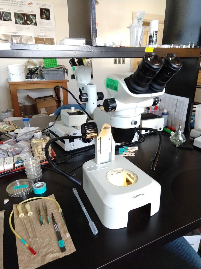 This is a photograph of my work station. This week I thought we could play a game of “guess that larvae!” I took the three pictures posted below, A, B and C. You tell me what common intertidal animals they come from and you win! Answers next week. A)  B)  C) 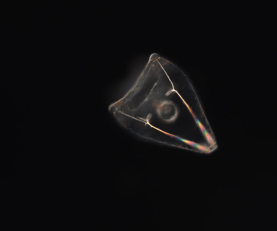 |

AuthorMy name is Philip Aspinall, and I am a student at Sierra College in Grass Valley, California. The first time I peered into a microscope and found an entire, complex, beautiful world below the visible, I was transfixed. I am thankful for George von Dassow and Svetlana Maslakova for allowing me to work in their lab, and to Geroge for his generosity with his time and for being my mentor this summer. Archives
August 2019
Categories |


 RSS Feed